Current Research
Causal Neural Circuits in Emotion Regulation
We investigate the neural circuits and therapeutic mechanisms for emotion regulation in patients with mood disorders and brain lesions, aiming to guide targeting optimization in noninvasive neuromodulatory treatment.
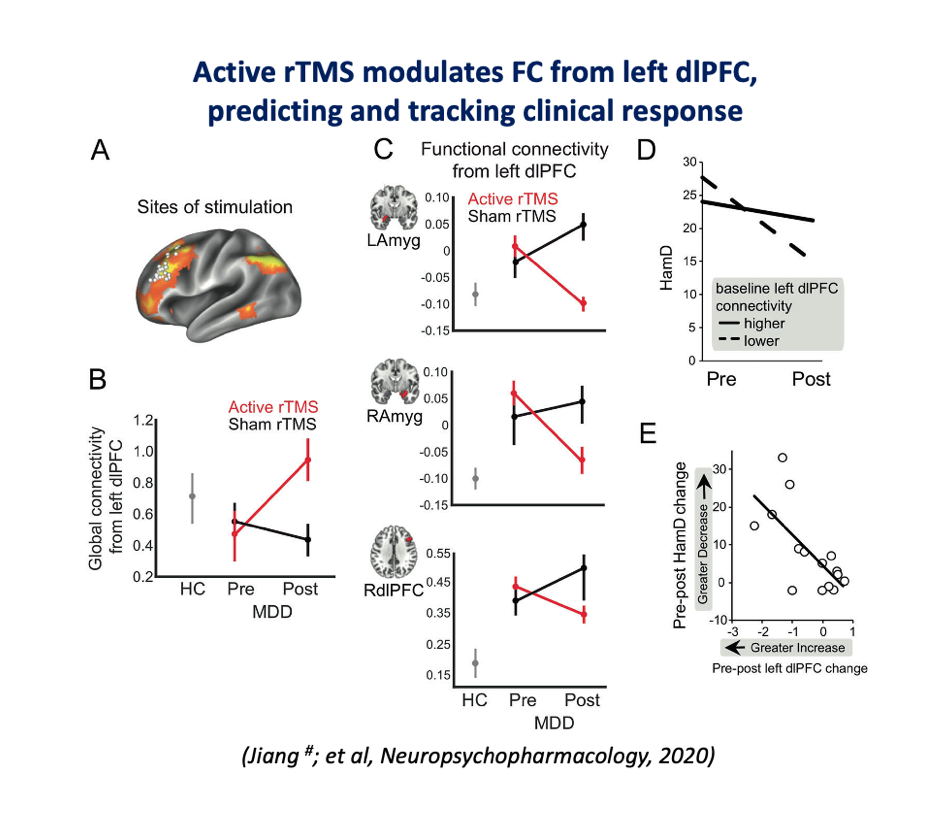
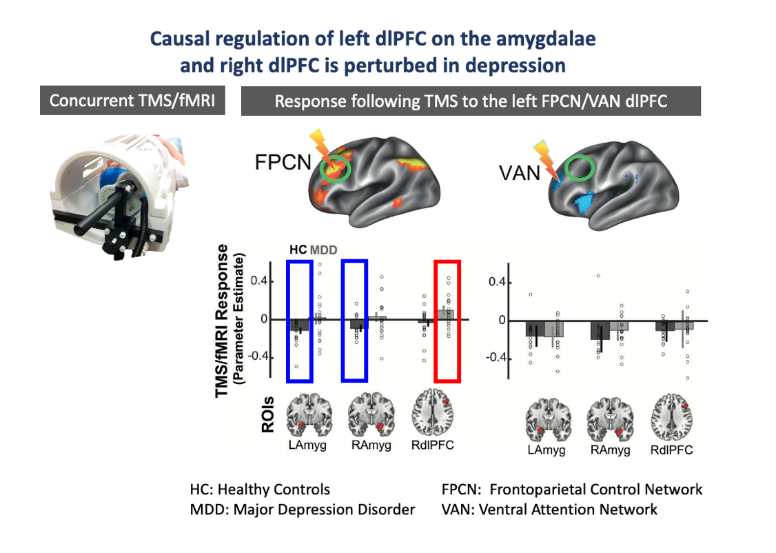
The Left Dorsolateral Prefrontal Cortex Causally Regulates Amygdala
Emotion regulation (ER) is a core affective ability that influences the quality of social interaction and is implicated in a variety of affective disorders. Evidence has shown that the left dorsolateral prefrontal cortex (dlPFC) is a key region involved in ER and an effective target for repetitive transcranial magnetic stimulation (rTMS) treatment in depression. Yet, our understanding of the causal regulatory role of the dlPFC and how rTMS to this region exerts its antidepressant effect is minimal. We performed a series of multimodal experiments in depression. Using concurrent TMS/fMRI, we observed an inhibitory effect of the dlPFC on the amygdala in healthy controls, but this effect was perturbed in depressed patients, indicating an altered dlPFC-amygdala circuit in depression. Our second experiment demonstrated this hypothesis by showing treatment with active but not sham rTMS to the dlPFC drove the dlPFC-amygdala connectivity pattern in patients towards to that seen in healthy controls. The findings together suggest that the rTMS may have improved the regulation of the dlPFC on the amygdala in depression, providing important clinical implications for treatment optimization.
- Jiang J. #, Eshel N. #, Keller C. #, Wu W. #, …, Etkin A. (2020). Global Connectivity and Local Excitability Changes Underlie Antidepressant Effects of Repetitive Transcranial Magnetic Stimulation. Neuropsychopharmacology, 45 (6): 1018-1025 (#: co-1st author). [Link]
|
|
Emotion Regulation Maps to a Network Centered on the Left Ventrolateral Prefrontal Cortex
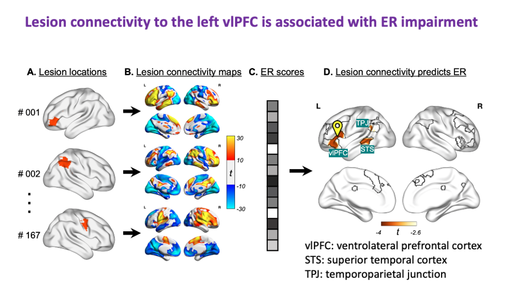
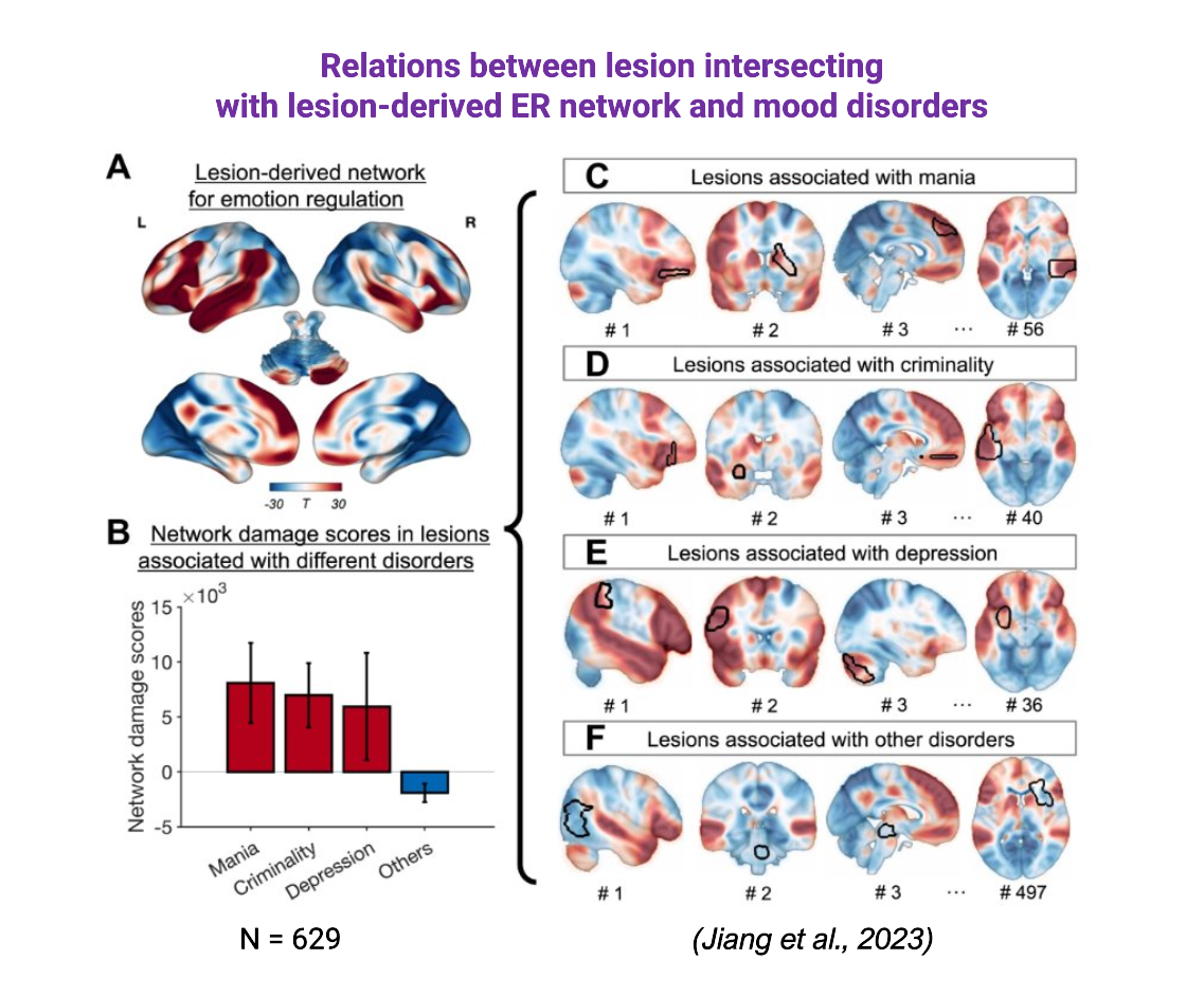
Lesions in multiple different brain locations have been associated with changes in emotion regulation (ER). However, it is unknown whether lesions causing ER impairment map to a connected brain network. We found that damage to these networks was associated with emotion regulation impairment in patients following focal brain injury (n = 167). Next, we used this lesion dataset to derive a de novo brain network for emotion regulation, which was defined by functional connectivity to the left ventrolateral prefrontal cortex (vlPFC). Finally, we used an independent lesion database (n = 629) to test whether damage to this lesion-derived network would increase the risk of neuropsychiatric conditions associated with emotion regulation impairment. We found that lesions causing mania, criminality, and depression intersected this network more than lesions causing other disorders. We conclude that emotion regulation maps to a connected brain network centered on the left vlPFC. Damage to this network impairs emotion regulation and may increase the risk of specific neuropsychiatric disorders.
- Jiang J., Ferguson M., Grafman J., Cohen A., & Fox M. (2023). A Lesion-Derived Brain Network for Emotion Regulation. Biological Psychiatry, 94(8), 640–649. [Link]
|
|
Invasive and Noninvasive Causal Evidence of Amygdala Engagement by DLPFC Stimulation
Invasive modulation of amygdala activity has shown promise in treating certain refractory psychiatric cases, but its widespread use among millions of treatment-resistant patients is impractical and entails inherent neurosurgery-related risks. Recent studies indicate that transcranial magnetic stimulation (TMS) of the dorsolateral prefrontal cortex (DLPFC) provides a potential noninvasive alternative. However, given the amygdala's deep location in the medial temporal lobe, there is a critical need for definitive causal evidence to determine whether and how DLPFC stimulation engages the amygdala. This study aims to obtain converging causal evidence of amygdala engagement to DLPFC stimulation using multimodal invasive and noninvasive imaging methods. This comprehensive approach seeks to elucidate the causal relationship between DLPFC stimulation and amygdala engagement, potentially informing noninvasive neuromodulatory therapies for psychiatric disorders.
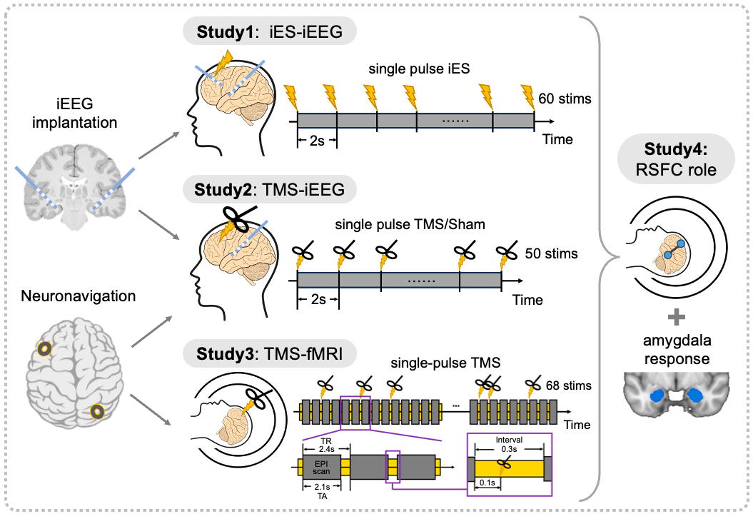
Causal Evidence of Hippocampus Engagement by Parietal Stimulation
The hippocampus is a key subcortical region essential for human memory and other cognitive functions, structurally and functionally connected to cortical regions in the parietal lobe. While evidence suggests that parietal lobe stimulation can influence memory performance, its precise causal modulation of hippocampal neural responses remains unclear. To address this, we employed advanced perturbation-measure methodologies combining invasive and non-invasive neural stimulation techniques, including transcranial magnetic stimulation (TMS) and intracranial electrical stimulation (iES), with concurrent neuroimaging approaches, including functional magnetic resonance imaging (fMRI) and intracranial electroencephalography (iEEG). This approach enabled the measurement of hippocampal responses evoked by parietal stimulation. We aim to unravel the causal connectivity within the parietal-hippocampus circuit, providing valuable insights into its functional dynamics and offering potential applications for developing non-invasive neuromodulatory therapies to enhance hippocampus-related cognitive functions in clinical populations.
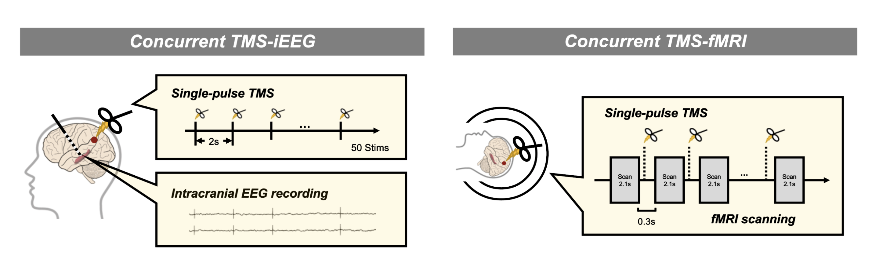
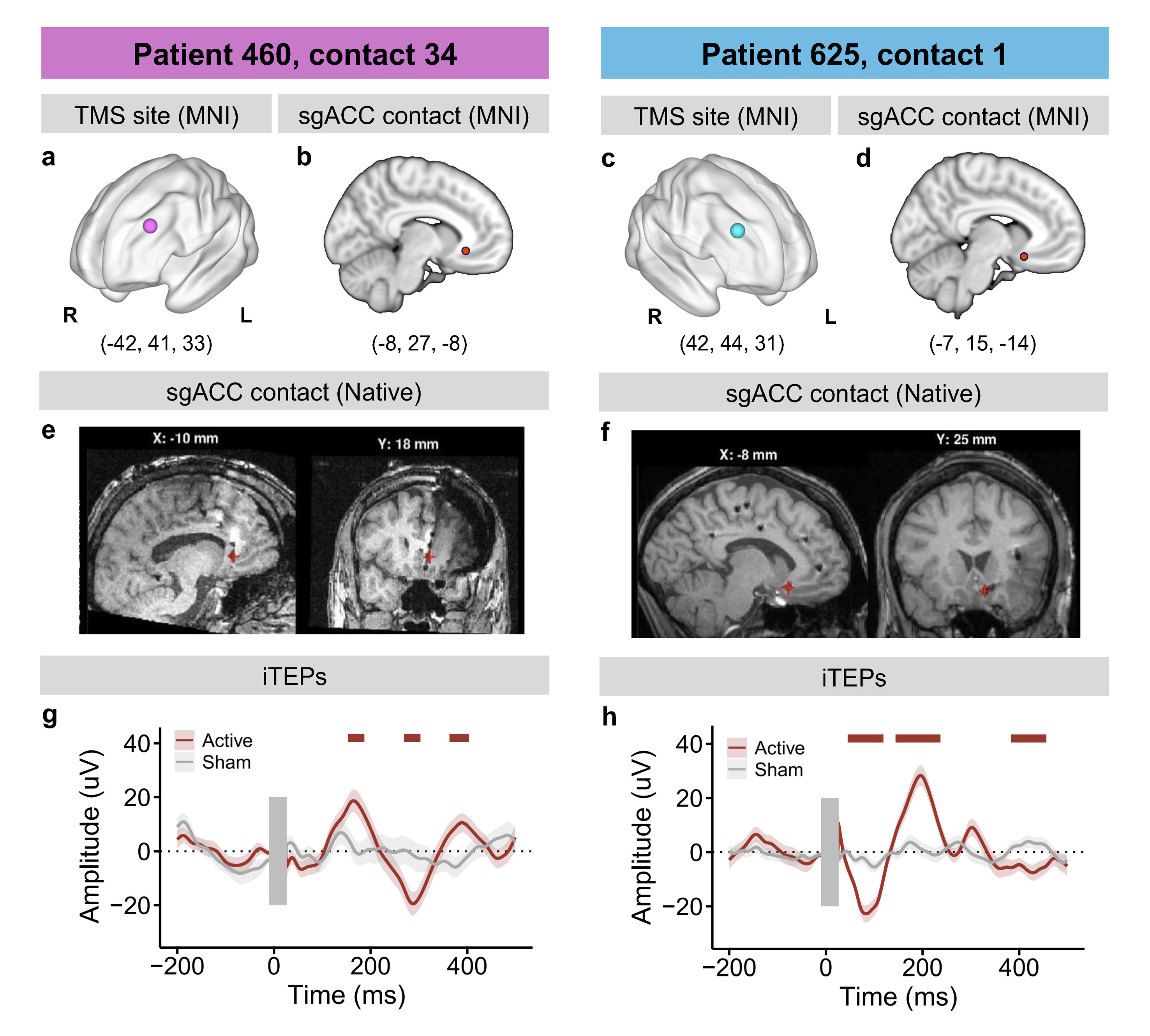
Dorsolateral Prefrontal Cortex TMS Evokes Responses in the Subgenual Anterior Cingulate Cortex: Intracranial EEG Evidence from Two Human Cases
The subgenual anterior cingulate cortex (sgACC) plays a key role in the pathophysiology of psychiatric disorders, particularly major depressive disorder, with its activity correlating with depression severity and treatment response, making it a promising target for neuromodulation. Meanwhile, transcranial magnetic stimulation (TMS) targeting the dorsolateral prefrontal cortex (dlPFC) has emerged as a noninvasive treatment for depression. A hypothesis suggests that TMS to the dlPFC may exert antidepressant effects by engaging the sgACC, which is typically anti-correlated with the dlPFC. This study explores the effects of dlPFC TMS on the sgACC using intracranial EEG in two human cases. The results show that single-pulse TMS applied to the dlPFC evokes significant responses in the sgACC, an effect not observed when TMS is applied to the parietal cortex in a control participant. Additionally, intracranial electrical stimulation of the dlPFC also produces sgACC responses. Resting-state fMRI connectivity analysis reveals an anti-correlation between the dlPFC and ACC regions, with individual dlPFC-to-sgACC connectivity weaker than normative values. Our findings underscore a functional relationship between the dlPFC and sgACC, offering new insights into how prefrontal stimulation may influence limbic regions involved in mood regulation.
Past Research
Social Interaction in Naturalistic Verbal Contexts
We study neural and cognitive mechanisms during dynamic and active social interaction in diverse naturalistic verbal contexts, and how the altered neural patterns reflect changes in non-verbal and verbal behaviors.
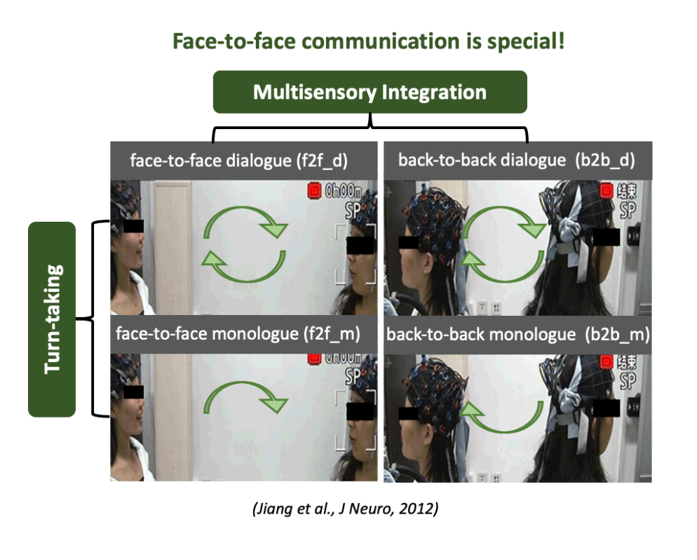
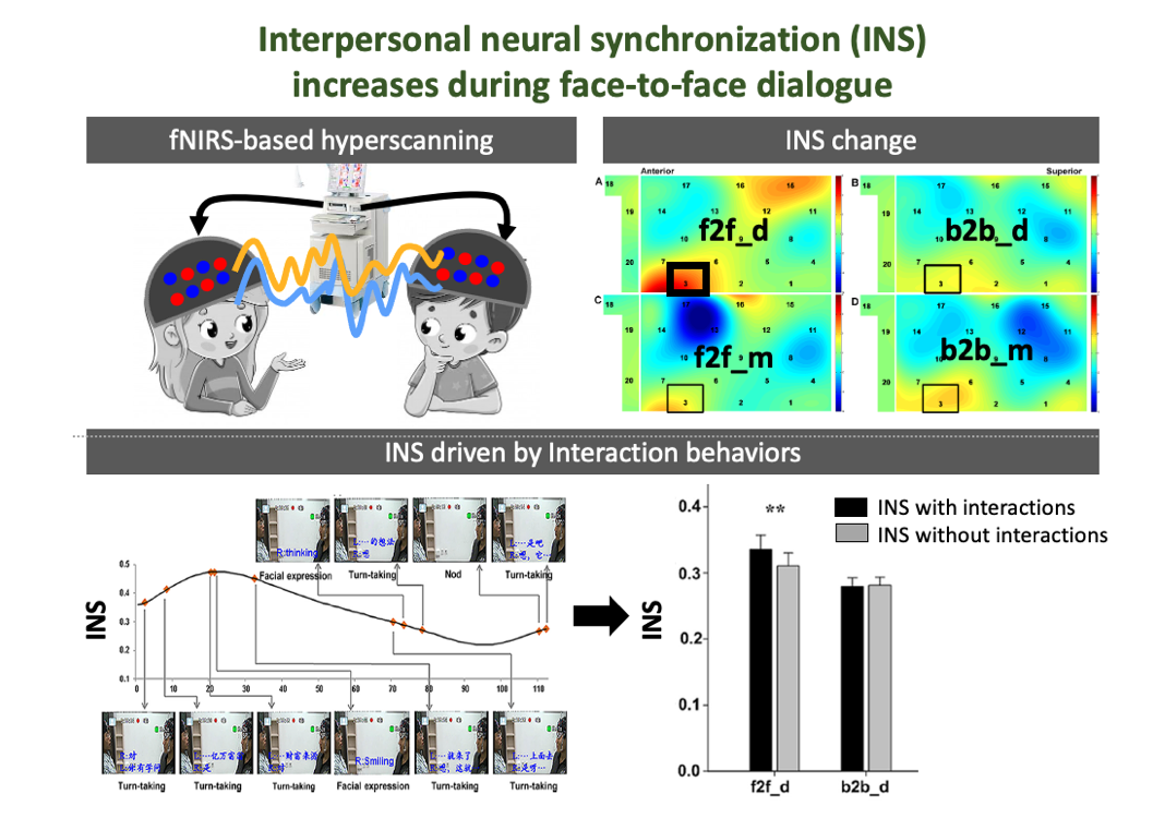
Face-to-face communication has unique neural features
Although the human brain may have evolutionarily adapted to face-to-face communication, other ways of communication, e.g., telephone and e-mail, now increasingly dominate our daily social interaction. However, the unique neural features of face-to-face communication relative to other types of communications remain unknown. We developed a functional near-infrared spectroscopy (fNIRS)-based hyperscanning to simultaneously measure activities from two communicating brains. We identified a significant interpersonal neural synchronization (INS) increase in the inferior frontal cortex during a face-to-face dialogue between communicating partners but not during other conditions (i.e., face-to-face monologue, back-to-back dialogue, and back-to-back monologue). Moreover, this INS increase resulted primarily from the multisensory integration such as facial expressions during the face-to-face dialogue. These results suggest that face-to-face communication, particularly dialog, has special neural features that other types of communication do not have. This study also contributes a naturalistic approach (fNIRS-based hyperscanning) and a novel neuromarker (INS) to the field of social neuroscience, catalyzing a new wave of research focusing on how multiple brains interact with each other under diverse face-to-face interaction settings.
- Jiang J., Dai B., Peng D., Zhu C., Liu L., Lu C. (2012). Neural synchronization during face-to-face communication. Journal of Neuroscience, 32(45):16064 –16069. [Link]
|
|
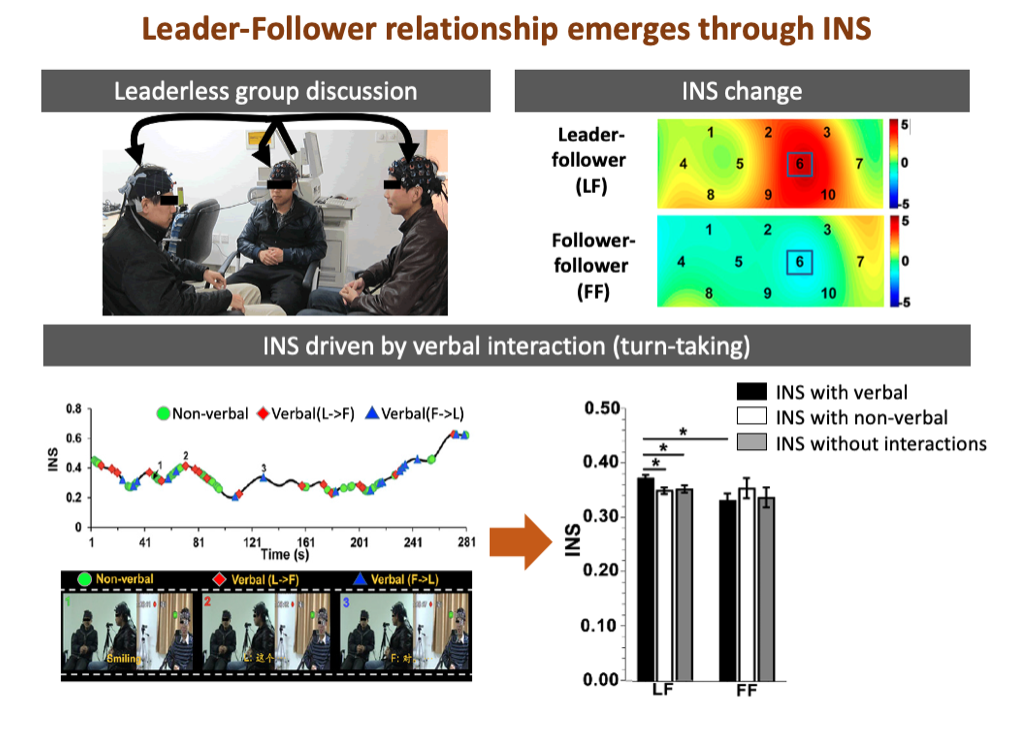
Humans build relationships through interpersonal neural synchronization (INS)
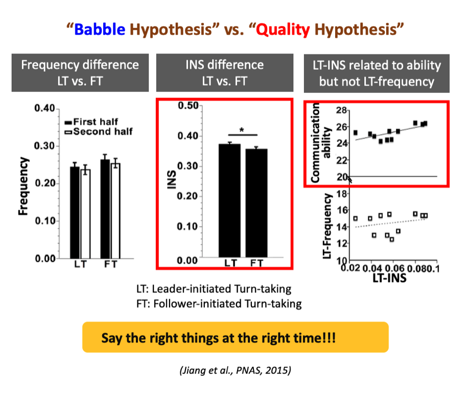
Human society consists of different interpersonal relationships. One important relationship in both evolutionary past and modern society is the leader-follower relationship. However, little is known about how the leader-follower relationship emerges through INS during social interaction. We leveraged fNIRS-based hyperscanning to simultaneously measure brain activities from three-person groups during a dynamic leaderless group discussion (LGD). We found a significantly higher INS in the left temporoparietal junction between leaders and followers than between followers. This difference was mainly driven by leader-initiated interactions and was related to leaders’ communication skills and competence, but not their communication frequency, in favor of the "communication quality hypothesis" in leader emergence. The INS even predicted when this relationship emerged along the course of LGD. These findings together suggest that humans may build new relationships with others by synchronizing their brain activities with others. Leader-follower relationship emerges because leaders are able to say the right things at the right time.
- Jiang J., Chen C., Dai B., Shi G., Liu L., Lu C. (2015). Leader emergence through interpersonal neural synchronization. Proceedings of the National Academy of Sciences of the United States of America, 112(14): 4274-4279. [Link]
|
|
A hierarchical model for interpersonal verbal communication
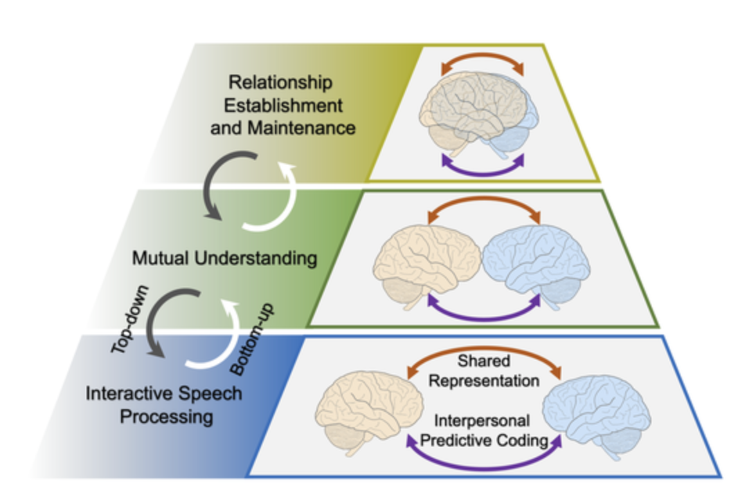
The ability to use language makes us human. For decades, researchers have been racking their minds to understand the relation between language and the human brain. Nevertheless, most previous neuroscientific research has investigated this issue from a ‘single-brain’ perspective, thus neglecting the nature of interpersonal communication through language. With the development of modern hyperscanning techniques, researchers have begun probing the neurocognitive processes underlying interpersonal verbal communication and have examined the involvement of interpersonal neural synchronization (INS) in communication. However, in most cases, the neurocognitive processes underlying INS are obscure. To tentatively address this issue, we propose herein a hierarchical model based on the findings from a growing amount of hyperscanning research. We suggest that three levels of neurocognitive processes are primarily involved in interpersonal verbal communication and are closely associated with distinctive patterns of INS. Different levels of these processes modulate each other bidirectionally. Furthermore, we argued that two processes (shared representation and interpersonal predictive coding) might coexist and work together at each level to facilitate successful interpersonal verbal communication. We hope this model will inspire further innovative research in several directions within the fields of social and cognitive neuroscience.
- Jiang J., Zheng L., Lu C. (2021). A hierarchical model for interpersonal verbal communication. Social Cognitive and Affective Neuroscience, invited Special Issue, 16(1-2): 246-255. [Link]
Altered neural patterns reflect changes in non-verbal and verbal behaviors
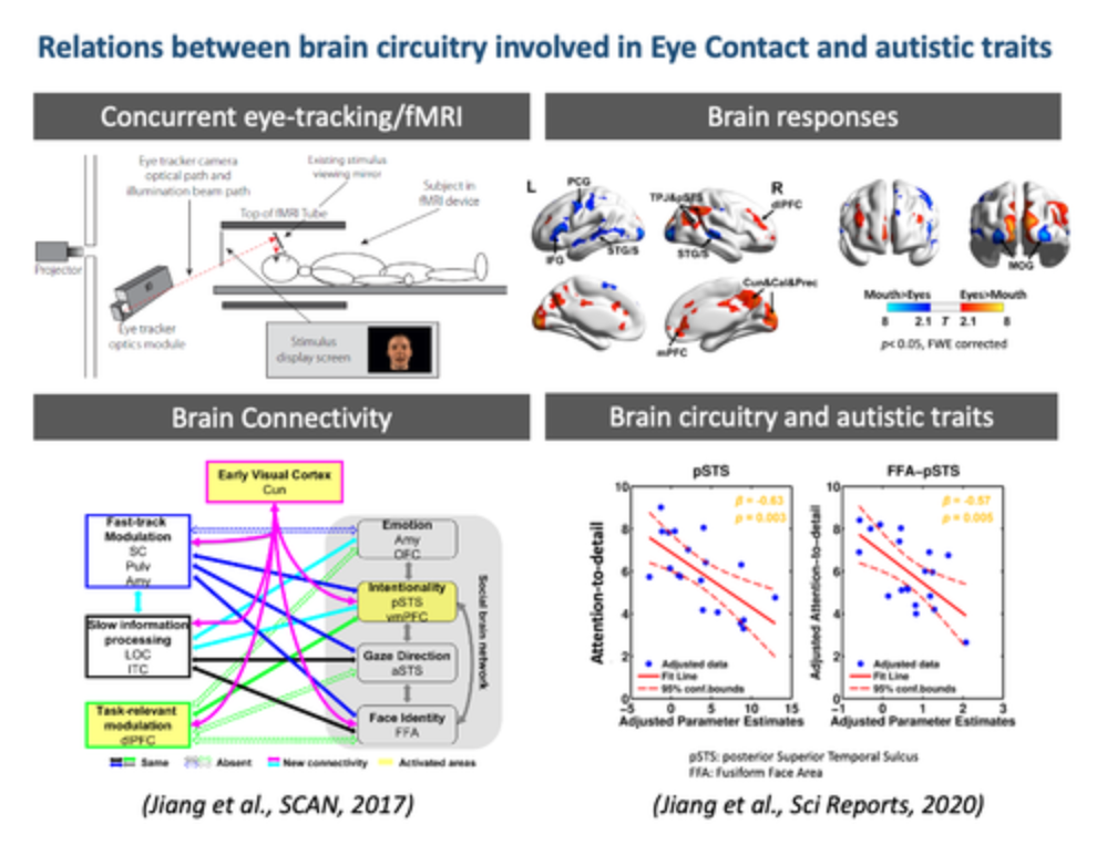
Eye contact is an important non-verbal cue for social interaction and occurs frequently and spontaneously in verbal communication. However, the brain circuitry underlying this behavior is unclear. We developed a novel paradigm to simulate eye contact in verbal communication. Using concurrent eye-tracking/fMRI, we simultaneously recorded participants’ eye movements and neural patterns while they were freely watching a pre-recorded speaker talking. We found an extensive network of brain regions showing specific responses and connectivity patterns to eye contact (Jiang et al., 2017, SCAN). Interestingly, the connectivity strength between brain areas involved in face processing was related to the level of autistic traits (Jiang et al., 2020, Scientific Reports).
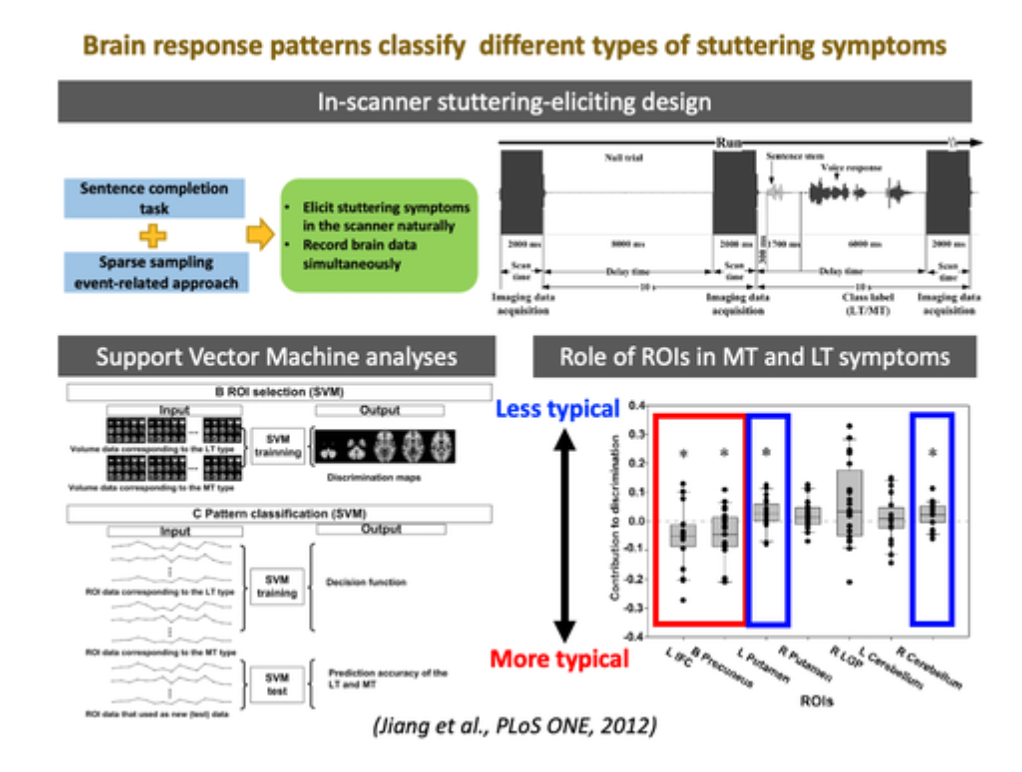
Like reduced eye contact, stuttering is a common communication difficulty that affects social interaction. Using a sparse-sampling fMRI paradigm, we designed a speech production task to naturally elicit patients’ stuttering symptoms in the scanner and simultaneously measure their brain responses to various stuttering symptoms. Unique patterns of altered brain activity were noted for different stuttering symptoms. The findings have important theoretical and clinical implications for patients with distinct dominated stuttering symptoms (Jiang et al., 2012, PLoS ONE).
- Jiang J., Borowiak K., Tudge L., Otto C., von Kriegstein K. (2017). Neural mechanisms of eye contact when listening to another person talking. Social Cognitive and Affective Neuroscience, 319–328. [Link]
- Jiang J., von Kriegstein K., Jiang J. (2020). Brain Mechanisms of Eye Contact during Verbal Communication Predict Autistic Traits in Neurotypical Individuals. Scientific Reports, 10 (1): 14602. [Link]
- Jiang J., Lu C., Peng D., Zhu C., Howell P. (2012). Classification of types of stuttering symptoms based on brain activity. PLoS ONE, 7(6): e39747. [Link]
|
|